Naevi are pigmented or non-pigmented skin tumors that contain nevus cells and are present in adults. Naevis can be elevated, flat, stalked, smooth, papillomatous, hair-bearing, or non-hair bearing. The most common naevi are classified as junctional or intradermal. A combination of these properties is called compound nevi.
The biggest challenge for the possible removal of lesions is to rule out sinister pathology. If a suspected lesion requires histological examination, the physician may recommend a biopsy of a pigmented lesion1. It is common for patients applying for cosmetic surgery to perform aesthetic removal of naevi along with various other benign lesions such as keratosis, dermatosis, papulosa nigra, DPN, sebaceous gland hyperplasia, syringoma, and verruca.
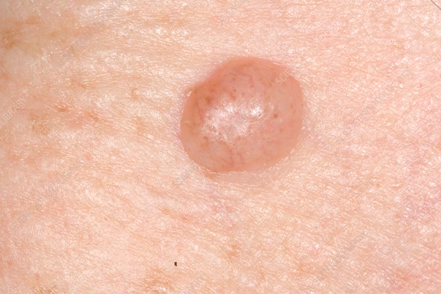
All forms of melanocytic naevus seem to have the same degree of pigmentation as the surrounding skin. Melanocytes do not pass on their pigmentation when they are present on the dermis or at epidermal transitions, as is the case with junctional naevi compounds. Only when the classic skin color of the moles is elevated above the skin surface do most people recognize them as such.
They appear to grow from pre-existing pigmented moles. They usually develop at the end of childhood and occur at some point in adulthood, although they are a rare new phenomenon after the age of 60. Most commonly affected are women aged 35 to 50.
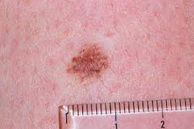
Most skin lesions with these properties are harmless and can be evaluated by experts by means of dermatoscopy. Short-term digital dermatoscopic imaging can be used for ambiguous or shallow lesions to monitor changes over time. If the suspicion persists, the melanoma can be removed by histopathology or diagnostic biopsy. A partial biopsy is not recommended as it can overlook areas with cancerous changes.
Our Intradermal Naevi Treatment Clinics are available in Central London, North London, Stanmore, Pinner, Rickmansworth, Watford etc.
Most of the intradermal or melanocytic naevi are harmless & can be left alone. But some may be required naevi removal treatment in the following circumstances.
A partial biopsy is not recommended as it can overlook areas with cancerous changes.
Naevi surgery procedures include:
300 Hampstead House
176 Finchley Road
London
NW3 6BT